Retinal Vein Occlusion
What is a retinal vein occlusion?
You can think of the retinal veins like the pipes that drain a sink. The blood that supplies the retina with oxygen drains from the retina through the retinal veins. If a retinal vein becomes occluded, the blood “backs up” and the pressure in the vein increases, much like water in a clogged drainage pipe. Unlike a steel pipe, however, blood can leak out of a vein that has a blockage. With nowhere else to go, the blood seeps into the retina. Retinal edema (swelling) occurs, and the vision becomes blurred. Retinal vein occlusions are commonly associated with high blood pressure.
Are there more than one type of retinal vein occlusion?
Yes. There are two main types of retinal vein occlusions: central and branch retinal vein occlusions.
How do the two types of retinal vein occlusion differ?
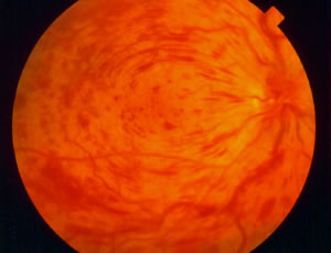
 A central retinal vein occlusion (CRVO), as its name implies, results from a blockage in the central retinal vein that drains all blood from the retina. Depending on the severity of the occlusion, the vision can range from quite good (without symptoms) to very poor. The photograph to the right is the typical appearance of a CRVO, with enlargement of the retinal veins and extensive blood throughout the retina:
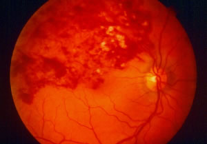
 A branch retinal vein occlusion (BRVO) is similar to a CRVO except that it only affects one branch of the central retinal vein. BRVO is more common than CRVO and typically does not cause as much visual loss, although the amount of visual loss can range from none to severe. To the left is an example of a BRVO, with blood involving one quadrant of the retina:
How are retinal vein occlusions treated?
Medications are frequently injected into the eye to reduce swelling in the retina and hopefully improve the vision. Avastin, Lucentis, Eylea and triamcinolone are the primary options for injection in patients with CRVO. The Ozurdex implant is a newer option for retinal vein occlusions. It is injected into the eye much like the above medications but delivers a steroid medication called dexamethasone in a sustained-release manner over three to six months.
When a CRVO is severe enough to cause a widespread lack of blood supply to the retina, abnormal blood vessels sometimes grow. Usually, they grow in the front part of the eye where fluid (not blood) normally drains from the eye. These abnormal vessels can block the drain, causing the pressure in the eye to become elevated. This is a type of glaucoma called “neovascular glaucoma”. It can cause severe pain and even more visual loss. Patients with CRVO are monitored closely for the growth of such blood vessels. If they are detected, Avastin or Lucentis can be injected into the eye and laser treatment can be performed to prevent them from causing the pressure in the eye to increase.
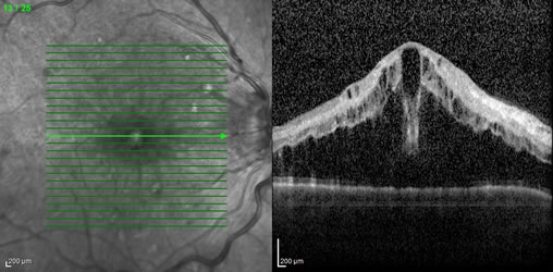
 There are more options for treatment of BRVO. Laser treatment can be performed in order to reduce swelling in the retina, and the vision can sometimes be improved with this treatment. As in the case of CRVO, Avastin, Lucentis and triamcinolone can be injected into the eye to reduce swelling in the retina and hopefully improve the vision. Also similar to CRVO, the Ozurdex implant is an option for treating retinal swelling due to BRVO. If the occlusion is severe enough to cause a lack of blood supply to the retina, abnormal blood vessels may grow. Usually, these vessels grow on the retinal surface in the case of BRVO. They can cause visual loss, usually by bleeding and less often by causing retinal detachment. Laser treatment can be performed to reduce the abnormal blood vessel growth and prevent visual loss related to bleeding or retinal detachment. On the right is an example of optical coherence tomography (OCT) showing the sort of swelling we typically see with a retinal vein occlusion:


