Retinal Tear/Detachment
What does the retina have in common with a camera or wallpaper?
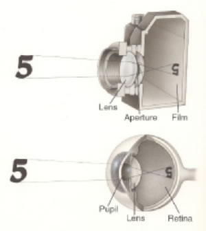 The eye is very similar in its structure to a camera. You can think of the retina as a small bit of photographic film. As the film captures images that can be made into photographs, the retina captures images and sends them to the brain to be interpreted as vision. The retina also needs to be flat against the inner wall of the eye in order to function properly, just as film needs to be flat inside the camera to take good photographs. The image to the right illustrates this comparison:
You can also think of the retina like wallpaper. It lines the inner wall of the back of the eye much like wallpaper lines a wall. Just as wallpaper would look strange if part or all of it were not attached to the wall, the retina needs to be completely attached to the inner lining of the eye in order to function well.
How does a retinal tear occur?
The vitreous is a clear gel that fills the back portion of the eye. When we are born, it is quite solid, like firm gelatin, and is in contact with the retina. You can picture the eye at birth like a hard-boiled egg; the vitreous is like the egg white (except it is clear), in contact with the retina much as the egg white is in contact with the egg shell. As years pass, the vitreous becomes more liquid and begins to pull away from the retina. Eventually, in most eyes, the vitreous separates from the retinal surface. This event often causes strange visual perceptions, sometimes described as cobwebs, flashes of light, and floaters. The cobwebs and flashes are probably caused by stimulation of the retina as the vitreous separates from it. The floaters usually represent the vitreous, itself, now visible as it floats in front of the retina. The following video provides additional information about “flashes and floaters”:
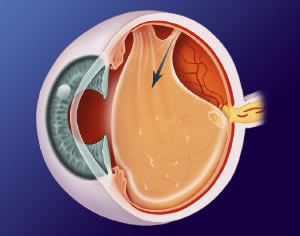 The separation of the vitreous from the retina is a normal event that usually has no harmful consequences. However, the vitreous is occasionally extra adherent to the retina in certain places, and as it separates from the retina, it sometimes takes some retinal tissue along with it. The result is a retinal tear. The picture to the left illustrates the separation of the vitreous from the retina that can result in a retinal tear:
How is a retinal tear treated?
The goal of treatment is to prevent a retinal detachment. This is done by creating an adhesive scar around the tear that will prevent fluid from going through the tear and under the retina. Remembering our analogy of the wallpaper, treatment of a retinal tear is similar to staplingaround a tear in the wall paper so that nobody can put his/her hand through the tear and pull the wallpaper off the wall.
One of two methods can be used to treat a retinal tear: laser or cryotherapy. Laser involves either placing a lens against the eye for focusing the laser beam or delivery of the laser from a headset, using a hand-held lens to focus the laser. Cryotherapy involves placing a freezing probe on the white of the eye in the location of the retinal tear. The probe causes a full-thickness freeze, through the white of the eye and the retina. Both of these procedures can be performed in the office with local anesthesia. In most cases, a retinal detachment can be prevented, but the retina sometimes detaches despite treatment of the retinal tear.
How does a retinal detachment occur?
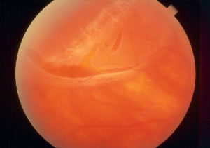 Returning to our analogy of the wallpaper, you can think of a retinal tear as if someone had cut a hole in the wallpaper. Theoretically, someone could then put his/her hand through the tear and pull the wallpaper off the wall. Similarly, once a retinal tear has developed, fluid from the vitreous can go through the tear and beneath the retina. The result is a retinal detachment. The photograph to the right shows multiple retinal tears with an associated detachment (a large, horizontal tear and a smaller, vertical tear; the retina is detached above and attached below):
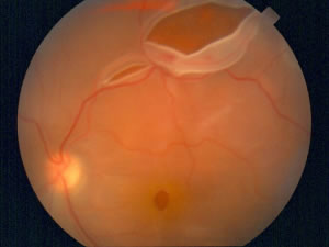 The photograph to the left shows a retinal detachment with a large retinal tear in the upper portion of the image and a smaller tear to the left of the larger one. This particular retinal detachment happens to be associated with a macular hole (the small, circular, dark area toward the bottom of the photograph):
How is a retinal detachment treated?
Again, think of the detached retina like wallpaper that has come off the wall. In order to re-attach the wallpaper, you could either 1) push the wallpaper back against the wall or 2) bring the wall in to the wallpaper. While option #2 does not make much sense when considering wallpaper, it is quite logical in the repair of retinal detachments.
Pneumatic retinopexy is the least invasive method of repairing a retinal detachment. It can be performed in the office under local anesthesia, without a trip to the operating room. It is a two-step procedure. First, cryopexy (as described above) is performed. Next, a gas bubble is injected into the eye through a very small needle. (In some cases, the gas is injected first and laser treatment is done later.) Following the procedure, the patient is usually asked to spend the majority of the time in a specific position so that the gas bubble covers the retinal tear, allowing the retina to reattach. Pneumatic retinopexy is only ideally suited for a subset of patients who have only one tear in the upper portion of the retina and have not yet had cataract surgery. The following video provides additional information about pneumatic retinopexy:
A scleral buckle is a soft silicone band that is place around the eye in order to indent the eyewall. It “brings the wall to the wallpaper,” so to speak. The silicone band is not visible to other people because it is located behind the eyelids. Over time, the eye forms a fibrous covering around the band, so it essentially becomes a part of the eye. Only in rare situations is it ever removed. In addition to placing the band around the eye, cryotherapy is performed, and fluid is sometimes drained from beneath the retina. Occasionally, a gas bubble is injected into the eye, as well. This procedure is performed in an operating room under local or general anesthesia. The following video provides additional information about the scleral buckle procedure:
A vitrectomy is the surgical equivalent of pushing the wallpaper back to the wall. It involves placing instruments through small holes in the white of the eye to remove the vitreous, drain the fluid from beneath the retina, apply laser treatment around any retinal tears, and place a gas bubble in the eye. Like a scleral buckle, this procedure is performed in the operating room under local or general anesthesia. Often following a vitrectomy, the patient is asked to maintain a certain head position for several days so that the gas bubble covers the retinal tear(s) as the laser scars make a firm adhesion around the tear(s). Vitrectomy is sometimes performed in conjunction with placement of a scleral buckle, at the surgeon’s discretion. The following video provides additional information about vitrectomy:

